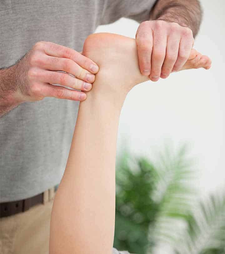
Achilles Pain – The Achilles tendon, the thickest and strongest tendon in the human body, is the combined tendon of the gastrocnemius and soleus muscles. Pain in the main body of the tendon (2-3cm above the insertion) appears to respond much better to treatment than pain at the insertion itself. The bursae are also important potential sources of pain, as is the superolateral tubercle of the calcaneum (heel bone), which, when excessively large, is called ‘Haglund’s Deformity’.
Achilles Tendinopathy
This type of injury to the Achilles tendon occurs when the load applied to the tendon, either in a single episode or over an extended period of time, exceeds the ability of the tendon to withstand the load. Factors that may predispose to Achilles tendinopathy include :-
1. Years of running
2. Increases in activity (distance, speed, gradient)
3. Decrease in recovery time between training sessions
4. Change of training surface
5. Change of footwear
6. Excessive pronation
7. Calf weakness
8. Poor muscle flexibility
9. Poor footwear
Clinical Features
Presentations can vary but typically include :-
Pain in morning and on activity, pain on stretching, tendon thickening, tenderness, redness, swelling.
Treatment of Mid-Portion Achilles Tendinopathy
Most evidence for treatment includes :-
1. Heel-drop exercises
2. GTN patches
3. Sclerosing injections
4. Shockwave therapy
5. Heel raises
Alfredson’s Heel-Drop Protocol (Eccentric Strengthening)
1. 3×15 repetitions, twice daily, 7 days per week, for 12 weeks
2. Do exercises with knee straight and knee bent (45 degrees)
3. Exercises are expected to be painful.
4. Progress exercises until they become pain-free.
5. Increase load / resistance until exercises are painful again.
6. Carry out a total of 180 drops/day.
It should be pointed out that certain groups of patients may not be able to tolerate the above protocol and it may have to be adapted on physiotherapist advice.
Glyceryl Trinitate (GTN) Patches
There is evidence that GTN patches, applied locally to the tendon, can reduce pain on activity by 12 weeks. The patches come in varying doses. A 0.5mg patch should be cut in quarters and applied to the site of maximum pain for 24 hours at a time and then replaced. A 0.2mg patch would best be cut in half and applied similarly.
Corticosteroid Injection
The injection of corticosteroids in Achilles tendinopathy has short-term pain relieving effects, but can be detrimental in the longer term without the appropriate period of rehabilitation.
Sclerosing Injections
This innovative treatment consists of using ultrasound guidance while injecting a vascular sclerosant (polidocanol) in the area of neovascularisation anterior to the tendon. Short- and long-term evaluation of this treatment has shown promising results and would require a specialist in lower limb disorders to carry out such a treatment. This can be arranged at the clinic.
Rehabilitation after sclerosing injection includes 1-3 days of rest, then gradually increased tendon loading activity, whilst being careful to avoid jumping, fast running and heavy strength training during the first 2 weeks. After this period, such activities that load the tendon maximally are permitted.
Electrotherapy Modalities
Treatments such as extracorporeal shock wave therapy and ultrasound can potentially improve the symptoms of Achilles tendinopathy in combination with an eccentric strengthening programme.
Adjunct Conservative Treatments
Biomechanical evaluation of the foot and leg is a clinically important part of Achilles tendon management and modification of foot posture in some patients can reduce pain and increase the capacity to load the tendon. Similarly, soft tissue therapy of the calf complex and tendon mobilisation, can assist in the overall management of Achilles tendinopathy.
Surgical Treatment
Procedures range from simple percutaneous tenotomy to removal of tendon pathology via an open procedure. Research has shown that 75% of patients reported good to excellent results after 18 months post-surgery. All Achilles tendon surgery requires early postoperative rehabilitation which needs to continue for 6-12 months. Wise patients will continue with a maintenance programme of physiotherapist-prescribed rehabilitation exercises even after having returned to training and competition.
Insertional Achilles Tendinopathy
Clinical Assessment
This condition is closely associated with retrocalcaneal bursitis and Haglund’s deformity. It is a condition of the ‘enthesis organ’. The physiotherapy assessment will include evaluation of the tendon, bursa and calcaneum with inspection of the region for bony prominence and local swelling as well as palpation of the area of maximal tenderness.
Ultrasound and MRI can help to assess the extent of pathology in the tendon and the bursa. Radiography can complement clinical assessment f the calcaneum and will reveal tendon calcification, if present.
Treatment
A heel lift worn inside both shoes is a good practical way of unloading the region. Alfredson’s heel drop protocol should be implemented into the rehabilitation programme, but it should be stated that it is less successful than when used for mid-portion tendinopathy.
Sclerosant injections with polidocanol have been shown to be effective. Additionally, symptoms can appear to arise from the retrocalcaneal bursa. In these cases the symptoms may respond to non-steroidal anti-inflammatory medication or intra-bursal corticosteroid injection. Abolition of pain after local anaesthesia helps confirm the diagnosis. Following injection, the patient should rest for 48 hours and then slowly resume activity, building up to full activity over a period of 2-3 weeks. If conservative management fails in cases of Haglund’s disease where a deformity is present, surgery is indicated.
Achilles Tendon Rupture
Complete rupture of the Achilles tendon classically occurs in athletes in their 30’s or 40’s. The typical patient is a 40 year old male, and the male : female ratio is 10 : 1. The patient describes a feeling “as if I was hit or kicked in the back of the leg” and pain is not always the strongest sensation. This is immediately followed by grossly diminished function. A snap or tear may be audible.
The patient will usually have an obvious limp but may have surprisingly good function through the use of compensatory muscles. That is, the patient may be able to walk, but not on the toes with any strength. Assessment and diagnosis are typically confirmed by the physiotherapist with the use of the Simmond’s or Thomson’s calf squeeze test. There may also be a palpable gap in the tendon.
Surgical Management
Surgical treatment of Achilles tendon rupture is associated with 27% lower risk of re-rupture compared with non-surgical treatment. Post-operatively, patients are placed in a functional cast or brace for up to 8 weeks. Because range of movement and strength can be difficult to regain after rupture repair, earliest possible mobilisation and rehabilitation is carried out by the physiotherapist.
Non-Surgical Management
Non-surgical management of an Achilles tendon rupture is not indicated for athletes, but may be appropriate for older patients or patients with a low level of activity. This involves cast immobilisation for up to 8 weeks with a structured rehabilitation programme.
Posterior Impingement Syndrome
This condition of the ankle refers to the impingement of the posterior talus by the adjacent aspect of the posterior aspect of the tibia in extremes of ankle plantarflexion. An enlarged posterior tubercle of the talus, or an os trigonum, may be present.
The os trigonum represents an unfused ossific centre in the posterior process of the talus. This is a normal anatomical variant present in approximately 10% of the population. The diagnosis of posterior impingement syndrome is suggested by pain and tenderness at the posterior aspect of the ankle and confirmed by a positive posterior impingement test.
If further confirmation is required, a small amount of a local anaesthetic can be injected around the posterior talus and the impingement test performed without pain. In practice, this is not always feasible and the test relies on the clinical accuracy of the physiotherapist.
Treatment
Treatment includes rest from exacerbating activities, manual mobilisation of the sub-talar, talo-crural and mid-foot joints, anti-inflammatory medication and electrotherapy modalities. If the condition persists, a corticosteroid injection around the area of maximal tenderness may reduce pain.
This is performed on the lateral side of the ankle, as the medial aspect contains the neurovascular bundle. Frequently this condition does not respond to conservative management and requires surgical removal of the posterior processor the os trigonum.
Sever’s Lesion
This is a common insertional enthesopathy among adolescents and is discussed earlier.
Accessory Soleus Muscle
Although considered a rare cause of Achilles region pain, anatomical studies suggest that an accessory soleus is present in about 6% of people. The typical presentation is pain and swelling and if symptoms do not settle, surgical removal of the accessory soleus may be the best treatment.
Other causes of pain in the Achilles region
Achilles bursitis is generally caused by excessive friction, such as by heel tabs, or by wearing footwear that is either too tight or too loose. Pressure can often be relieved by widening the heel of the boot manually or providing ‘donut’ protection to the area of bursitis as it resolves.