Anterior Thigh Pain – The two most important aspects of the history of a patient with anterior thigh pain are the exact site of the pain and the mechanism of injury. The site of the pain is usually well localised in cases of contusion or muscle strain. A contusion is likely to be the result of a direct blow, whereas a muscle strain usually occurs when an athlete is striving for extra running speed or kicking distance. Bilateral thigh pain usually suggests the pain is referred from the lumbar spine.
In anterior thigh pain of acute onset, the diagnosis is usually straightforward and examination is confined primarily to local structures. With symptoms of insidious onset, diagnosis is more difficult and examination includes sites that refer pain to the thigh, such as the lumbar spine, sacro-iliac joint and hip.
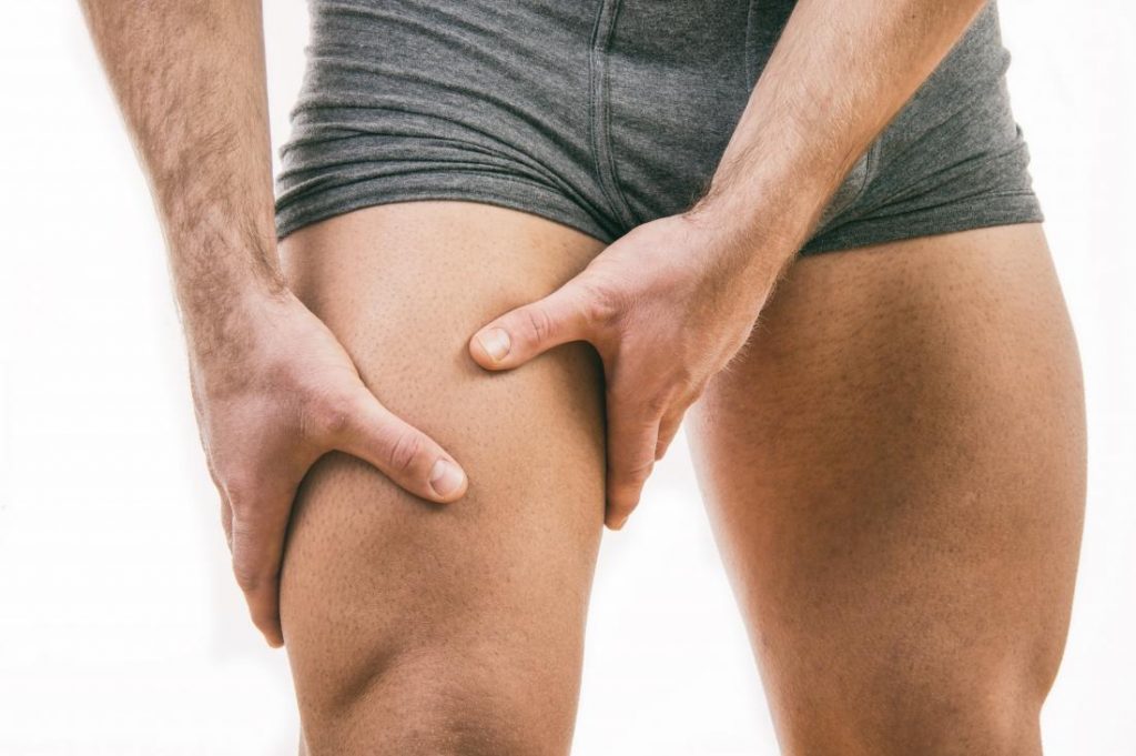
Quadriceps Contusion
If the patient reports a direct blow to the anterior thigh and examination confirms an area of tenderness and swelling with worsening pain on muscle contraction and stretch, thigh contusion with resultant haematoma is the most likely diagnosis.
The contusion may be either intramuscular or intermuscular. In the intermuscular haematoma, the blood escapes through the fascia and is distributed between the compartments of the thigh. The intramuscular haematoma is more painful and restrictive. Usually only a single quadriceps muscle will be affected.
Treatment
The treatment of thigh contusion can be divided into 4 stages :-
Stage 1 – Control of bleeding (RICE protocol, gentle stretch)
Stage 2 – Restoration of pain-free range of movement (Soft tissue therapy, static muscle contractions, stretching, ultrasound, exercise bike, swimming);
Stage 3 – Functional rehabilitation (Soft tissue therapy, increase stretch and strengthening exercises, squats, step downs, jogging)
Stage 4 – Graduated return to normal activity (As above, increase weight resistance, plyometrics, sports specific skills)
The most important period in the treatment of a thigh contusion is in the first 24 hours following the injury. The patient should carry out the RICE regimen immediately. The importance of rest, cold therapy and elevation is crucial at this stage. Depending on the seriousness of the injury, the use of crutches can be utilised to partially weight-bear.
In the acute management of a thigh contusion, ice should be applied in a position of maximal pain-free quadriceps stretch. The patient must be careful not to aggravate the bleeding by excessive activity, alcohol ingestion or the application of heat. The patient must be careful not to over-stretch as the bleed can recur.
Soft tissue therapy (massage) is contraindicated for the first 48 hours. After this, soft tissue therapy is aimed to promote lymphatic drainage, with the aim of avoiding bleeding to recur.
Compartment Syndrome of the Thigh
Intramuscular haematoma of the thigh after a blunt contusion may result in high intra-compartmental pressure of the thigh. Treatment of this condition is the same as that described above.
Myositis Ossificans
Occasionally after a thigh contusion, the haematoma calcifies. This condition can be seen on x-ray after 3 weeks following injury. If not diagnosed, after 6-7 weeks, a bony lump within the thigh musculature is often palpable.
Symptoms
An increase in morning pain and pain on activity. Pain at night is also common. On palpation, the developing myositis ossificans has a characteristic “woody” feel. Range of movement of the thigh on stretching is also restricted.
Treatment
Treatment may include local electrotherapy to reduce the muscle spasm and gentle, passive range of motion exercises. Evidence suggests that surgery is unhelpful for this condition and corticosteroid injection is absolutely contraindicated.
Quadriceps Muscle Strain
Strains of the quadriceps muscle usually occur during sprinting, jumping or kicking. Strains are seen in all the quadriceps muscles but are most common in the rectus femoris, which is more vulnerable as it passes over 2 joints : the hip and the knee.
Like all muscle strains, quadriceps strains may be graded into mild (grade 1), moderate (grade 2) or severe (grade 3). The patient feels the injury as a sudden pain in the anterior thigh during an activity requiring explosive muscle contraction. There is local pain and tenderness and if the strain is severe, swelling and bruising.
Grade 1 strain is a minor injury with pain on resisted active muscle contraction and on passive stretching. An area of local muscle spasm is palpable at the site of pain.
Grade 2 strains cause significant pain on passive stretching as well as active muscle contraction. There is usually a moderate area of inflammation surrounding a tender palpable lesion.
Grade 3 tears or complete ruptures occur with a sudden onset of pain and disability during intense activity. A muscle fibre defect is usually palpable when the muscle is contracted.
Treatment
It is important that the athlete regains pain-free range of motion as soon as possible. Loss of strength will be more marked than a thigh contusion injury and there is a strong emphasis on strength retraining.
The rehabilitation programme should commence with low resistance, high repetition exercise. Concentric and eccentric exercises should begin with very low weights. General fitness can be maintained by activities such as swimming and upper body training. Functional retraining should be incorporated as soon as possible.
Proximal Rectus Femoris Strains
This injury has also been termed the “bull’s eye lesion”, and occurs within the belly of the muscle as opposed to the more common muscle-tendon junction. The patient typically complains of a tender anterior thigh mass and weakness and/or pain with activities such as running and kicking.
Recent evidence has shown that an average return to full training in professional footballers after a comprehensive rehabilitation programme was 27 days compared to just 9 days for peripheral rectus femoris strains.
Differentiating between a mild quadriceps strain and a quadriceps contusion
Occasionally, it may be difficult to distinguish between a minor contusion and a minor muscle strain. A general rule is that a patient with a thigh strain should progress more slowly through a rehabilitation programme, than someone with a diagnosed muscle contusion.
The patient with a thigh strain should also avoid sharp acceleration and deceleration movements in the early stages of injury. So, all you footballers, tennis players, squash players, rugby players etc etc have been warned !!!
Stress Fracture of the Femur
Although this condition is uncommon, it should be suspected in an athlete, especially a distance runner, who complains of a general non-specific dull ache in the anterior thigh. Pain may also be referred to the knee. There may be tenderness over the shaft of the femur that can be aggravated if the patient sits with the leg hanging over the edge of a bench. This is known as the “hang test” or ” fulcrum test”.
Treatment
Treatment involves rest from painful activities and maintenance of fitness carrying out non-weight bearing exercises such as cycling or swimming. When the “hang test” is completely negative, on average after seven weeks, it is thougght to be safe to return to normal sporting activities on a gradual basis.
Referred Pain
Referred pain may arise from the hip joint, the sacro-iliac joint or the lumbar spine. Patients with referred pain may not have a history of injury and have few signs suggesting local injury. An increase in neural tension may suggest that referred pain is a contributing factor. The Modified Thomas’s test is the most specific neural tension test for a patient with anterior thigh pain.
If the modified Thomas’s test reproduces the patient’s anterior thigh pain and altering the neural tension (passive knee flexion/extension) affects the pain, the lumbar spine and psoas muscle should be examined carefully.
Commonly there is hypomobility of the upper lumbar intervertebral joints on the affected side. Mobilisation of these joints will often significantly reduce symptoms. Deep soft tissue treatment to the psoas muscle may also be effective.