Rear Foot Pain
Foot Pain – The rear foot consists of the calcaneus and talus bones. The most common cause of rear foot pain is plantar fasciitis, although fat pad contusion and stress fractures of the calcaneus can occur.
Plantar Fasciitis
The plantar aponeurosis is composed of 3 segments, all arising from the calcaneus. The central, and clinically most important segment arises from the plantar aspect of the posteromedial calcaneal tuberosity and inserts into the toes to form the longitudinal arch of the foot.
The aponeurosis provides static support for the longitudinal arch and dynamic shock absorption. Plantar fasciitis is essentially an overuse condition of the plantar fascia at its attachment to the calcaneus.
Causes
Individuals with pes planus (low arches or flat feet) or pes cavus (high arches) are at increased risk of developing plantar fasciitis. The condition commonly results from activities that require maximal plantarflexion of the ankle and simultaneous dorsiflexion of the metatarsophalangeal joints (e.g. running or dancing). In the older patient, it may be related to excessive walking in inappropriate or non-supportive footwear. Reduced ankle dorsiflexion, obesity and work-related weight-bearing are additional independent risk factors for its development.
Finally, and importantly, plantar fasciitis is commonly associated with tightness in the proximal myofascial structures, especially the calf, hamstring and gluteal regions, due to the continuous nature of fascia throughout the lower limb.
Clinical Features
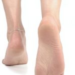
The pain is usually of gradual onset and felt classically on the medial aspect of the heel. Initially it is worse in the morning and decreases with activity, often to ache post-activity. As the condition becomes more severe, the pain may be present when weight-bearing and worsen with activity.
Examination reveals acute tenderness along the medial tuberosity of the calcaneus, and may extend some centimetres along the medial border of the plantar fascia. The plantar fascia is generally tight and stretching may reproduce pain.
Investigations
Referral for further investigations may be necessary.
Ultrasound is the gold standard diagnostic investigation, with swelling of the plantar fascia the typical feature. X-rays are often performed and may show a calcaneal spur, but are not essential for diagnosis.
Treatment
1. Avoidance of aggravating activity
2. Ice treatment after activity
3. Anti-inflammatory medication
4. Stretching of plantar fascia, gastrocnemius / soleus complex
5. Self-massage with bottle or golf ball
6. Strengthening exercises of intrinsic muscles of the foot
7. Taping
8. Silicone gel heel pad
9. Night splints
10. Soft tissue therapy
11. Corticosteroid injection
12. Shockwave therapy
13. Surgery
Fat Pad Contusion
The fat pad acts as a shock absorber, protecting the calcaneus at heel strike.
Causes
This may develop either acutely after a fall onto the heels from a height, or chronically because of excessive heel strike with poor heel cushioning.
Clinical Features
The patient often complains of marked heel pain, particularly during weight-bearing activities. The pain is often felt laterally in the heel due to the pattern of heel strike. Examination reveals tenderness, often in the posterolateral heel region. There may also be an area of redness.
Treatment
Consists primarily of avoiding aggravating activities, particularly excessive weight-bearing. Ice therapy and anti-inflammatory medication can also be effective in the acute stages. As the pain settles, the use of a silicone gel heel pad and good footwear are important as the athlete resumes activity. Heel lock taping will often provide symptomatic relief.
Midfoot Pain
The midfoot is comprised of the 3 cuneiform bones, the cuboid and the navicular bones, as well as the surrounding soft tissues. The most common cause of midfoot pain is midtarsal joint sprain after ankle injury, but the most important cause is a stress fracture of the navicular bone.
Stress Fracture of the Navicular
These are amongst the most common stress fractures seen in the athlete, especially in sports that involve sprinting, jumping or hurdling.
Cause
A combination of overuse and training errors plays a significant role in the development of navicular stress fractures.
Clinical Features
The onset of symptoms is usually insidious, consisting of a poorly localised midfoot ache associated with activity. The pain typically radiates along the medial aspect of the longitudinal arch and the symptoms abate rapidly with rest.
Examination reveals localised tenderness at the ‘N’ spot, located at the proximal dorsal portion of the navicular.If palpation confirms tenderness over the ‘N’ spot, the athlete should be considered to have a navicular stress fracture until proven otherwise.
Treatment
Treatment of a navicular stress reaction (no cortical breach) is weight-bearing rest, often in an air cast, until symptoms and signs have resolved, followed by a graduated return to activity. Treatment of navicular stress fracture is strict non-weight-bearing immobilisation in a cast for 6-8 weeks.
At the end of this period the cast should be removed and the ‘N’ spot palpated for tenderness. Generally, the ‘N’ spot will be non-tender but if tenderness is present, the patient should have the cast reapplied for a further 2 weeks of non-weight-bearing immobilisation. Management must be based on the clinical assessment as there is poor CT and MRI correlation with clinical union of the stress fracture.
Some clinicians advocate surgical treatment with the insertion of a screw in cases where there is significant separation of the fracture.
Post-Cast Rehabilitation and Prevention of Recurrence
Following removal of the cast, it is essential to mobilise the stiff ankle, subtalar and midtarsal joints. The calf muscles require soft tissue therapy and exercise to regain strength. These must be done before resuming running. Activity must begin gradually, slowly building up to full training over a period of 6 weeks.
Midtarsal Joint Sprains
The midtarsal joint (Chopart’s joint) consists of the talonavicular and calcaneocuboid joints. Other joints in the midtarsal area are the naviculocuneiform, cuboid cuneiform and intercuneiform joints. Injuries to the midtarsal joints are most commonly seen in gymnasts, jumpers and footballers.
Lisfranc’s Joint Injuries
This refers to the tarsometatarsal joints – the bases of the 5 metatarsals, with their corresponding 3 cuneiforms and cuboid bones. The spectrum of injuries of the Lisfranc’s joint complex ranges from partial sprains with no displacement to complete tears with separation (diastasis) of the 1st and 2nd metatarsal bones, and depending on the severity, different patterns of tarsal and metatarsal displacement.
Lisfranc’s Joint Fracture – Dislocation
There are 2 main mechanisms of injury :-
1. Direct
This is more uncommon and occurs as a simple crush injury to the tarsometatarsal joint region. There is no specific pattern of damage or distinctive appearance with a direct injury.
2. Indirect
This is more common and generally occurs secondary to a longitudinal force sustained while the foot is plantarflexed and slightly rotated. The extent of the damage depends on the severity of the injury : in milder injuries the weak dorsal tarsometatarsal ligaments are ruptured, while with more severe injuries, there may also be fractures of the plantar aspect of the metatarsal base, or the plantar capsule may rupture and the metatarsal may displace dorsally.
Clinical Features
A patient with this injury may complain of midfoot pain and difficulty weight-bearing, following an acute injury by the mechanisms described above. Pain is classically aggravated by forefoot weight-bearing, with the patient unable to run on his or her toes, and will feel pain on the push-off phase of running and sometimes during walking and on calf raises.
Midfoot pain that persists for more than 5 days post-injury should raise suspicion of a Lisfranc’s joint injury.
Examination
1. Tenderness with or without swelling on the dorsal midfoot, often with associated bruising in this region
2. Pain with combined eversion and abduction of the forefoot while the calcaneus is held still.
Investigations
Referral for further investigation may be necessary.
Plain x-rays while weight-bearing are recommended. Diastasis between the 1st and 2nd metatarsal bases of greater than 2mm suggests a Lisfranc’s joint injury.
MRI scans have been shown to be sensitive in detecting tears of the Lisfranc’s ligament when plain x-rays appear normal and should be performed if there is a possible midfoot injury.
Treatment
Treatment depends on the degree of instability present. In grade 1 injuries, when there is no instability, conservative management is recommended with non-weight-bearing in a cast or air cast for 6 weeks. Following removal of the cast, mobilisation of the ankle and a calf strengthening programme are required. Orthoses may be needed to correct the intrinsic alignment of the foot and to support the 2nd metatarsal base. A graded return to activity is required.
If there is evidence of instability present, as in grade 2 and 3 injuries, surgical reduction and fixation is required. This is a significant injury that has a much better prognosis if managed correctly initially rather than being salvaged once there is prolonged joint malalignment and non-union. A delay in diagnosis has been associated with a poor outcome, a prolonged absence from sport, and chronic disability due to ligamentous instability of the tarsometatarsal joint.
Cuboid Syndrome
Peroneal tendinopathy is often associated with the development of the cuboid syndrome. Due to excessive traction of the peroneus longus, the cuboid becomes subluxated. Pain is experienced with lateral weight-bearing. There may also be a history of an inversion sprain. Most patients with this syndrome have excessively pronated feet but it is also seen in patients with lateral instability.
The peroneus longus tendon subluxates the cuboid bone so that the lateral aspect is rotated dorsally and medially. There may be a visible depression over the dorsal aspect of the cuboid. Treatment involves a single manipulation to reverse the subluxation. The cuboid is pushed upward and laterally from the medial plantar aspect of the cuboid.
Forefoot Pain
Forefoot problems range from corns, calluses and nail problems to bone and joint abnormalities, which may result from overuse and thus have an insidious onset.
Stress Fractures of the Metatarsals
The most common metatarsal stress fracture is at the neck of the 2nd metatarsal. This occurs in the pronating foot, when the first ray is dorsiflexed, resulting in the 2nd metatarsal being subjected to greater load. The 2nd metatarsal is also susceptible to stress fracture in the case of a Morton’s foot, where the first ray is shorter than the second. The base of the 2nd metatarsal is firmly fixed in position next to the cuneiform bones, further increasing the likelihood of fracture, which is common amongst ballet dancers.
Clinical Features
The patient with a metatarsal stress fracture complains of forefoot pain aggravated by activity such as running or dancing. The pain is not severe initially, but gradually worsens with activity. Examination reveals the presence of focal tenderness overlying the metatarsal.
Investigations
If the x-ray is negative, an isolated bone scan or MRI may confirm the diagnosis.
Treatment
The management of most stress fracture is straight-forward, involving rest from weight-bearing aggravating activities for approximately 4 weeks. If the patient is required to be on his or her feet excessively, the use of an air cast may be required for 1-2 weeks until pain settles. The patient should be allowed to recommence activity when he or she does not experience pain when walking and there is no local tenderness at the fracture site.
A graduated exercise programme should be instituted to return the athlete to full training and competition. Orthoses may be required to control abnormal foot mechanics.
Fractures of the 5th Metatarsal
Three different fractures affect the 5th metatarsal. The fracture of the tuberosity at the base of the 5th metatarsal is usually an avulsion injury that results from an acute ankle sprain. This uncomplicated fracture heals well with a short period of immobilisation for pain relief.
A serious fracture of the 5th metatarsal is the fracture of the diaphysis, known as a Jones fracture. This may be the result of an inversion, plantarflexion injury, or more commonly, as a result of overuse. A Jones fracture requires 6-8 weeks of non-weight-bearing cast immobilisation, or in situations when rapid return to activity is required, immediate surgical fixation with the percutaneous insertion of a screw. Non-union may be treated by bone grafting or screw fixation.
More recently, there has been a tendency to favour early screw fixation due to concerns regarding the high incidence of failure of cast treatment. In one study, early screw fixation resulted in quicker times to union and return to sport compared to cast treatment. The average time to return to sport after this procedure appears to be approximately 8 weeks, although early return to sport may predispose the athlete to re-fracture, and it may be wise to wait for full radiographic healing before return to sport.
An acute spiral fracture of the distal third of the 5th metatarsal is seen, especially in dancers who suffer this fracture when they lose their balance on demi pointe and roll over the outer border of the foot. Undisplaced fractures of this type may be treated with weight-bearing rest, while displaced fractures may require 4-6 weeks of cast immobilisation.
Metatarsophalangeal Joint Synovitis
This condition is commonly referred to as “metatarsalgia”, which is an inflammatory condition occurring most frequently in the 2nd, 3rd and/or 4th metatarsophalangeal joints, or isolated in the first metatarsophalangeal joints.
Causes
The joints become inflamed due to excessive pressure over a prolonged period. It is often related to either, pes cavus or high arched foot, excessive pronation, prominent metatarsal heads or Morton’s foot.
Clinical Features
The patient complains of pain aggravated by forefoot weight-bearing, particularly in the mid-stance and propulsion phases of walking. The pain is usually gradual in onset.
Examination reveals local tenderness on palpation and the pain is aggravated by passive forced flexion of the toe. It most commonly affects the 2nd metatarsophalangeal joint, followed by the 1st and 3rd joints. There may also be an associated skin lesion (e.g. callus) over the plantar surface of the affected joint due to the excessive load.
Investigations
Referral for an X-ray may be necessary to assess the degree of degeneration of the joint.
Treatment
Treatment requires appropriate padding to redistribute weight from the painful areas together with footwear that has adequate midsole cushioning. Anti-inflammatory medication may be helpful and occasionally corticosteroid injection is required.
First Metatarsophalangeal Joint Sprain (Turf Toe)
This is a common injury occurring in athletes in which the plantar capsule and ligament of the 1st metatarsophalangeal joint is damaged.
Causes
The classic mechanism of injury is usually that of a forced hyperextension of the 1st metatarsophalangeal joint, although occasionally a plantarflexion injury to the joint may result in this injury. Predisposing risk factor include; competing or training on artificial turf, pes planus or excessive pronation and soft, flexible footwear.
Clinical Features
The athlete usually complains of localised pain, swelling and occasionally redness at the 1st metatarsophalangeal joint following a ‘bending’ injury to the joint. The pain is classically aggravated by weight-bearing or movement of the big toe.
Examination reveals localised swelling and tenderness at the 1st metatarsophalangeal joint. Passive plantarflexion and dorsiflexion of the 1st metatarsophalangeal joint are generally painful, and there may be a reduction in the range of movement in both directions.
Treatment
Treatment consists of ice, anti-inflammatory medication, electrotherapy modalities and decreased weight-bearing for at least 72 hours. Additional treatment may include taping and the use of stiff-soled shoes the 1st metatarsophalangeal joint from further injury. Recovery generally takes 3-4 weeks.
A possible long-term sequel to this injury is the development of hallux limitus.
Hallux Limitus
This condition is defined as a restriction in dorsiflexion of the hallux at the 1st metatarsophalangeal joint, secondary to exostoses or osteoarthritis of the joint.
Causes
1. Trauma – secondary to chondral damage
2. Excessive pronation of the foot may increase the stresses on the joint
3. Repetitive weight-bearing
4. Rheumatoid arthritis
5. Hypermobile first ray
Clinical Features
The main presenting symptom is usually that of pain around the 1st metatarsophalangeal joint. The pain is often described as a deep aching sensation that is aggravated by walking, especially in high heels, or activities involving forefoot weight-bearing.
In patients with longstanding hallux limitus, a distinct shoe wear pattern is seen : the sole demonstrates wear beneath the 2nd metatarsophalangeal joint and the 1st interphalangeal joint.
Examination reveals tenderness of the 1st metatarsophalangeal joint, especially over the dorsal aspect, often with palpable exostoses. There is a painful limitation of joint motion with the degree of limitation reflecting the severity of the arthrosis.
Investigations
It may be necessary to refer for an X-ray. Plain X-rays display the classic characteristics of degenerative osteoarthritis and the degree of degeneration observed will reflect the duration and severity of the condition.
Treatment
Conservative management consists of an initial reduction in activity, anti-inflammatory medication, a cortisone injection if required, physiotherapy, biomechanical correction involving orthoses within footwear, and if these measures fail, surgery would be indicated.
Hallux Valgus
This condition is defined as a static subluxation of the 1st metatarsophalangeal joint. It is characterised by lateral deviation of the great toe and medial deviation of the first metatarsal. Bony exostoses develop around the 1st metatarsophalangeal joint, often with an overlying bursitis. In severe cases, exostoses limit 1st metatarsophalangeal joint range of motion and cause pain with the pressure of footwear.
Causes
1. Constricting footwear
2. Excessive pronation – increased pressure on the medial border of the hallux.
Clinical Features
As the deformity develops, pain over the medial eminence occurs. The pain is typically relieved by removing the shoes or by wearing soft, flexible, wide-toes shoes. Blistering of the skin or development of an inflamed bursa over the medial eminence may occur.
Investigation
Plain x-rays should be performed to assess both the severity of the deformity and the degree of 1st metatarsophalangeal joint degeneration.
Treatment
Initial treatment involves appropriate padding and footwear to reduce friction over the medial eminence. Correction of foot function with orthoses is essential. In more severe cases, surgery may be required to reconstruct the 1st metatarsophalangeal joint and remove the bony exostoses. Orthoses are often required after surgery.
Sesamoid Injuries
The 1st metatarsophalangeal joint is characterized by the two sesamoid bones which play a significant part in the function of the great toe.
Causes
The sesamoid bones may be injured by traumatic fracture or sprain of the sesamoid-metatarsal articulation. Sesamoid abnormality involves inflammatory changes and osteonecrosis around the sesamoid. The medial sesamoid is usually affected. Inflammation may be caused by landing after a jump, increased forefoot weight-bearing activities (e.g. sprinting and dancing) or after traumatic dorsiflexion of the hallux.
Clinical Features
The patient complains of pain with forefoot weight-bearing and will often walk with weight laterally to compensate. Examination reveals marked local tenderness and swelling overlying the medial or lateral sesamoid. Movement of the 1st metatarsophalangeal joint is usually painful and often restricted. Resisted plantarflexion of the great toe elicits both pain and weakness.
Treatment
Treatment of sesamoid inflammation is with ice, anti-inflammatory medication and electrotherapy modalities to reduce inflammation. Padding can also distribute weight away from the sesamoid bones.
Corticosteroid injection into the joint space between the sesamoid and metatarsal may prove effective if underlying abnormalities have been corrected. Orthoses are required if foot mechanics are abnormal.
In elite athletes, such as basketballers and tennis players, treatment of sesamoid fractures, involves up to 6 weeks of non-weight-bearing in an air cast or short leg cast.
Freiberg’s Osteochondritis
This condition affects adolescents between the ages of 14 and 18 years. The metatarsal head appears fragmented on x-ray. Offloading of the metatarsal heads using padding and orthoses is essential to prevent permanent metatarsal head flattening that may predispose to adult osteoarthritis.
Joplin’s Neuritis
This condition involves compression and irritation of the dorsal medial cutaneous nerve over the 1st metatarsal and 1st metatarsophalangeal joint. It usually occurs because of irritation from footwear.
The patient complains of pain radiating along the first ray into the hallux. Wearing appropriate footwear and using foam and felt to redistribute the load from the affected area generally provides relief. Orthoses may be required to prevent excessive pronation.
Morton’s Neuroma
This condition is a swelling of nerve and scar tissue arising from compression of the interdigital nerve, usually between the 3rd and 4th metatarsals. The patient complains of pain radiating into the toes, often associated with pins and needles and numbness. Pain is increased by forefoot weight-bearing activities and with narrow-fitting footwear.
Examination reveals localised tenderness. Excessive pronation contributes to metatarsal hypermobility and impingement of the interdigital nerve.
Treatment consists initially of ice to alleviate acute tenderness. Plantar metatarsal padding is used to spread the load over the metatarsals. Intrinsic foot muscle strengthening exercises are indicated to maintain or improve the transverse arch. However, in chronic cases, little improvement is seen with padding.
Injection of corticosteroid and local anaesthetic agents in conjunction with the padding may provide lasting relief. The use of orthoses is essential if excessive pronation is present. If the patient obtains no relief, surgical excision of the damaged nerve is indicated.
Corns and Calluses
In the feet, corns and calluses result from uneven weight distribution and thus indicate abnormal foot biomechanics or poorly fitting footwear. Treatment involves their removal with a scalpel, the wearing of well-fitting footwear and, if abnormal foot mechanics are present, orthoses. Petroleum jelly over the corn or callus and on the outside of the sock can also help.
Ingrown Toenail
This arises from abnormal nail growth, trauma or poor nail cutting. Patients often present in acute pain with tenderness on gentle palpation. Nails are often infected. Treatment with local and oral antibiotic therapy is required. Conservative treatment involves cutting the offending nail and removing as much as possible.
If this does not relieve the pain then surgical intervention may be necessary. This is done using local analgesia, the problem nail is removed and a chemical is applied to the nail bed to permanently destroy the nail bed and prevent the nail ever regrowing.