Hip & Groin Pain – The most common causes of acute hip / groin pain are strains of the adductor muscles, iliopsoas muscles, or injuries to the hip joint itself, such as a labral tear and/or chondral injury.
Adductor Muscle Strains
Adductor muscle strains are a common injury in sports that involve sudden changes of direction. Examination findings are usually, localised tenderness, pain on passive abduction and pain on resisted adduction or combined flexion/adduction.
A typical treatment regimen consists of :-
(i) 0-48 hours – RICE protocol, active pain-free exercises
(ii) After 48 hours – depending on the size / grade of the muscle tear, gradually increase strengthening of abduction and adduction movement patterns using therabands, pulleys, light weights. Start stability exercises (e.g. pulleys with other leg, one-leg squats).
(iii) After 7-21 days – Functional strengthening, including cycling, pool running, jogging, swimming
(iv) After 21 days – Sport-specific drills, including running (straight line), running (figure of eight), rapid changes of direction, kicking (different forces and skills of kicking).
The clinical features of impingement tend to be pain on most shoulder movements and activities above 90 degrees elevation and in sports people during overhead activities such as throwing or tennis serving.
Treatment is aimed at settling down the symptoms through soft tissue techniques, electrotherapy, taping, exercises and postural education or corticosteroid injection.
Recurrent Adductor Muscle Strain
If not adequately rehabilitated of the initial injury, or resuming sport too quickly, these injuries can lead to chronic exercise-related groin pain.
In these cases, extra rehabilitation time through the stages should be provided, together with a detailed analysis of sporting technique to decide if their are any technical abnormalities affecting the rate of progress.
Acute Adductor Tendinopathy
Adductor tendinopathy causes proximal groin pain, which has a tendency to develop with increasing activity. If the condition remains untreated, the pain tends to persist during activity and may migrate either to the contralateral groin or to the suprapubic region.
Examination findings include local tenderness over the adductor origin and over the pubic tubercle, with pain on passive hip abduction and resisted hip adduction.
Treatment of an acute presentation includes a few days relative rest from the aggravating activity, followed by a gradual progression of adductor strengthening.
Iliopsoas Strains
The iliopsoas muscle is the strongest flexor of the hip joint. Iliopsoas problems may occur as an overuse injury resulting from excessive hip flexion, such as kicking. They present as a poorly localised ache that patients usually describe as being a deep ache in one side of the groin.
It is important to examine the lumbar spine as there is frequently an association between iliopsoas tightness and stiffness of the lumbar spine from which the muscle originates.
Treatment consists of avoiding aggravating activities, stretching of the psoas muscle and strengthening involving resisted hip flexion exercises. Often, mobilisation of the lumbar intervertebral joints at the origin of the iliopsoas muscles will result in an improvement of symptoms.
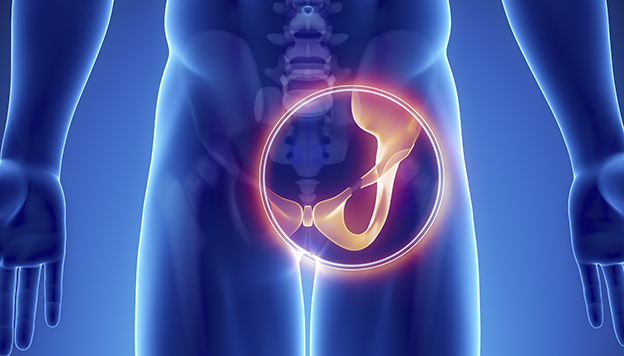
Hip Joint Injuries
Pain from the hip joint may be felt in a number of areas, including the hip, groin, outer thigh and lower back. The pain is commonly a dull ache , although it may be associated with a clicking or catching sensation. The pain may also be referred to the anterior aspect of the knee.
The hip may be inflamed as a generalised rheumatological disorder. Osteoarthritis of the hip may also been seen in the older patient. The pain of the osteoarthritic hip may be worse in the mornings or after activity.
In children between 5 and 12 years, Perthes’ disease should be considered, while in older adolescents between the ages of 12 and 16 years, a slipped capital femoral epiphysis may cause hip pain. A young patient with a slipped capital femoral epiphysis may present with very litle pain, but with a painless limp.
Examination
There are a number of tests to assess irritation and restriction of the hip joint. These include : the circumduction test; hip quadrant flexion, adduction and internal rotation; internal rotation with added adduction; flexion, abduction and external rotation (FABER or Patrick’s Test); and the impingement test.
It is unusual to find a case of an injured hip that does not have coexisting iliopsoas dysfunction. The probable causes of this presentation are due to direct irritation of the iliopsoas as it crosses the anterior aspect of the hip joint; and the iliopsoas being overloaded as a hip flexor due to restriction in hip movement. Examination findings would normally include; myofascial tightness in the iliopsoas muscle on abdominal palpation; and restricted hip extension on modified Thomas testing.
Investigations
Included are plain radiographs (X-rays) of the pelvis and lateral views of the hip. A full pelvis view is necessary to be able to assess the hip joint for the presence of dysplasia and to compare the two sides.
Evidence of dysplasia includes a short or angled roof, and signs of acute-chronic impingement, such as an os acetabulae, femoral bossing, Ganz lesions and impingement cysts. A ‘Ganz lesion’ is a thickening or bump on the superior femoral neck in response to chronic impingement in this area. The thickening of the bone then causes worsening impingement and a vicious cycle develops. The thickened bone often needs to be surgically resected.
MRI scanning of the hip to detect any intra-articular pathology is also useful. Injection of the hip joint with local anaesthetic with post-injection assessment of pain is very accurate by examining the hip pre- and post-injection.
Arthroscopy of the hip joint remains the most sensitive and specific investigation. In patients presenting with pain in the areas described, the clinician should maintain a high index of suspicion for labral injury, irrespective of the results of investigative imaging.
Synovitis
Synovitis is a complication of most hip injuries but can present as a primary problem, particularly when associated with rheumatological conditions. It usually responds well to X-ray guided injections of corticosteroid.
Labral Tears
With the increasing use of MRI and hip arthroscopy, labral tears of the hip joint are being increasingly recognised as a cause of hip pain. The mechanism of injury can be due to extrinsic factors (road traffic accident, lifting/twisting incident, repetitive squatting anh hip flexing) or intrinsic factors (dysplasis, Ganz lesions, torn ligamentum teres).
The natural history of an untreated acetabular labral tear is slow but progressive degeneration to degenerative hip joint disease. Treatment of these injuries involves arthroscopic debridement of the torn part of the labrum, which generally produces good results, especially in those with minimal or no associated chondral damage.
Trochanteric Bursitis
This can be due to bursitis in one of several bursae around the hip and it is aggravated by activities involving regular hip movements, such as climbing stairs and getting out of a car.
There are two bursae around the greater trochanter. The gluteus medius bursa and the trochanteric bursa. Gluteus medius tendinopathy and bursitis often exist together. The site of tenderness in these conditions is typically immediately above the greater trochanter and pain can be reproduced on stretching the gluteus medius.
Treatment for these conditions involves initial rest from aggravating activities, stretching and strengthening of the gluteus medius. Corticosteroid injection may be required and should be placed into the area of maximal tenderness above and behind the greater trochanter. As the condition is often associated with biomechanical abnormality, muscle tightness or excessive lateral tilt of the pelvis, pelvic stability exercises may play an important role in treatment.
Specific Concerns for Adolescents
A number of specific conditions may cause acute hip and groin pain in adolescents. Conditions of the hip joint such as Perthes’ disease, a slipped capital femoral epiphysis and epiphyseal fractures are major concerns.
Acute avulsion fractures and chronic apophysitis occur in adolescents as a result of excessive muscle contraction. The most common site is the attachment of the long head of the rectus femoris from the anterior inferior iliac spine.
Avulsion fractures present as an acute onset of anterior hip pain with a marked loss of function. On examination, there is localised tenderness and restriction of movement. In most cases, these should be treated conservatively as for a severe rectus femoris strain. Surgical reattachment of the avulsed fragment is usually not necessary but may be required when the fragment is separated from the bone by greater than 3cm. Rehabilitation is required for up to three months in these cases.